Overview
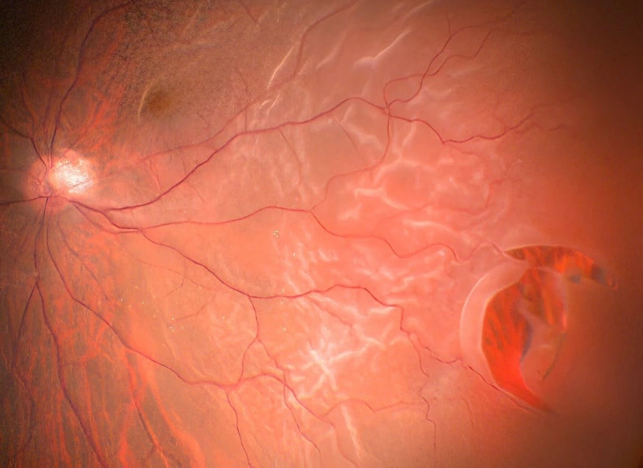
Retinal detachment occurs when the fine layer of tissue at the back part of the eye peels away (detaches) from its original position.
Signs of Retinal Detachment
Common signs of retinal detachment, although symptoms may vary in each case, include the following:
- The appearance of specks that obscure the vision
- Light flashes affect one or both eyes, a condition known as photopsia.
- Hazy vision
- Peripheral or side vision worsens.
If you notice signs of vision deterioration and suspect retinal detachment, it's crucial that you see your eye doctor immediately. Retinal detachment is considered a medical emergency and requires prompt treatment. Your quick action could make a significant difference in preserving your vision.
Causes of Retinal Detachment
- Rhegmatogenous detachment is a form of retinal detachment that occurs when a hole in the retina causes fluid to seep behind it. The accumulation of fluid can cause the retina to separate from its underlying tissue.
- Another reason is the formation of scar tissue on the surface of the retina, which causes the retina to detach from the back part of the eye.
- Retinal detachment can also occur from fluid build-up underneath the retina despite its absence of holes or tears.
Possible Risk Factors of Retinal Detachment
- Ageing is a potential risk factor, increasing the risk for those between forty and seventy.
- Having suffered a previous retinal detachment
- There’s a history of retinal detachment in the family
- Previous eye surgery like cataract removal
- A previous traumatic eye injury
Treatment for Retinal Detachment
Surgery is often considered when there’s a retinal hole, tear or detachment. Surgical techniques vary in each case. Dr Yako will discuss treatment options in detail, assessing your condition and eligibility thoroughly. We prioritise your care and comfort when treating something as serious as retinal detachment. Although considered a medical emergency, there is treatment available to help put you at ease.

Retinal Detachment Ultrasound

Retinal Detachment Schematic
Retinal Detachment
Overview
Iris repair is a surgical procedure that uses various techniques and materials, one of which is sutures, to restore the iris and pupil to their original, circular shape. This repair is necessary when the iris, a crucial part of the eye, is significantly damaged due to congenital abnormalities, traumatic eye injuries, or previous eye surgeries, causing distress to our patients.
Iris repair helps restore function to the iris after significant loss or damage, which may result in light sensitivity, glare, and accompanying visual disturbances such as blurred vision, double vision, and difficulty in focusing. Therefore, iris reconstruction focuses on returning the function of the iris as well as its anatomical appearance.
Surgical Techniques for Iris Repair
- The Use of Sutures: Sutures are versatile medical tools used for a variety of surgical procedures. In iris repair, sutures are used to re-contour or reconnect segments of the iris, particularly in the event of iris detachment (iridodialysis), with the aim of forming a round, anatomically appropriate pupil.
- McCannel Technique: The McCannel technique is a highly specialised surgical technique for repairing iris lacerations. It relies on sutures to secure the peripheral iris to the scleral wall.
- Iridodialysis Repair: This meticulous, well-known surgical approach involves the use of sutures to reconnect the iris to the ciliary body. In some cases, both an intraocular lens and a synthetic iris are used to repair the defect.
- Prosthetic Irises: Prosthetic irises are medical devices used for expansive iris defects. Depending on the patient's preferences and eligibility, there are several different prosthetic iris options, such as an iris lens diaphragm and synthetic, customizable iris implants. This variety ensures that patients can make informed decisions about their treatment.
Factors to Consider for a Successful Outcome
- The size and extent of the iris defect influence treatment options.
- Take into account ocular co-morbidities
- The patient’s preferences, such as cosmetic goals and functional needs, are considered.