Overview
Retinal vein occlusion (RVO) is an eye disorder that occurs when the vein in the retina, which is light-sensitive tissue found at the back part of the eye, suffers a blockage, significantly increasing the risk of severe vision deterioration and potential loss. When a vein in the retina is blocked, we refer to it as occlusion. Retinal vein occlusion may be due to a blood clot or when a more expansive blood vessel presses against the vein. With a degenerative disorder like retinal vein occlusion, the blood vessels weaken and leak. As a result, the macula thickens or begins to swell. Macular oedema results in reduced, severely poor, hazy vision.
This is just the beginning of a set of cataclysmic events because, with retinal vein occlusion, the blood vessels may develop within the iris, the colourised part of the eye. This abnormal formation increases pressure inside the eye, resulting in neovascular glaucoma. Abnormal blood vessels may also begin to develop in the retina, which increases the risk of bleeding at the back part of the eye and retinal detachment. This serious medical condition necessitates immediate and urgent medical attention to prevent further complications.
Primarily, there are two main forms of retinal vein occlusion such as:
- Central Retinal Vein Occlusion (CRVO) occurs when the main vein in the eye is obstructed.
- Branch Retinal Vein Occlusion (BRVO) occurs due to occlusion of the tiny blood vessels connected to the main vein of the eye.
Potential Risk Factors for Retinal Vein Occlusion
There are potential factors like underlying medical conditions that place you at higher risk of retinal vein occlusion, such as glaucoma, hypertension or chronic high blood pressure, vascular disease, obesity and diabetes.
Common Signs of Retinal Vein Occlusion
- Poor vision
- Hazy vision
- Eye floaters
- Eye pain is due to the formation of new blood vessels that increase pressure in the eye.
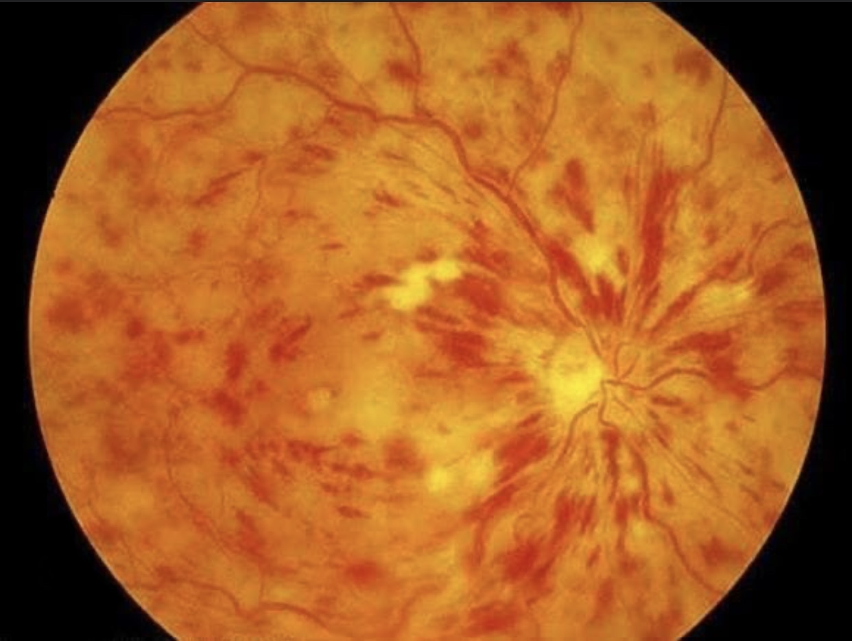
Retinal Vein Occlusion
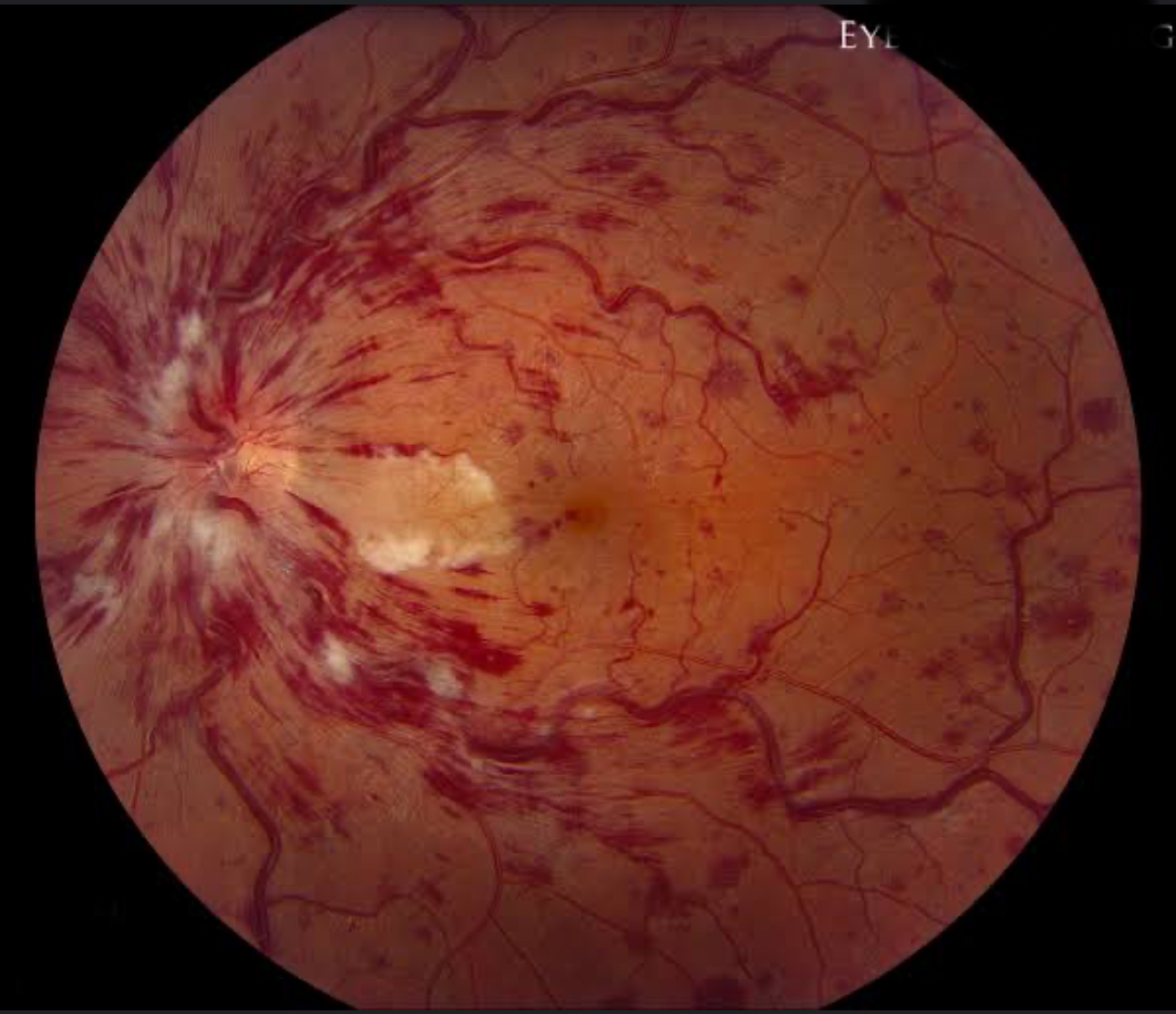
Retinal Vein Occlusion
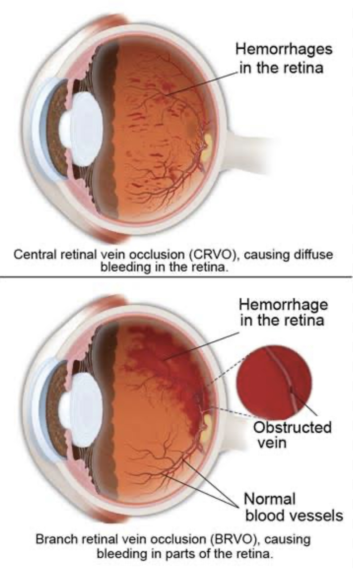
Retinal Vein Occlusion